Anatomy Of Chest Area - honors anatomy unit 5 lesson 5 - muscles of the abdomen ... : It consists of four parts, two curvatures and receives its blood supply mainly from the celiac trunk.
Anatomy Of Chest Area - honors anatomy unit 5 lesson 5 - muscles of the abdomen ... : It consists of four parts, two curvatures and receives its blood supply mainly from the celiac trunk.. The chest exam is performed more frequently than any other exam in the imaging department. Ct anatomy of the chest, axial reconstruction. Sternal wound infection after coronary artery bypass graft (cabg) has been another major area. Or motion attempt to minimize overlying osseous structures area of interest closest to image receptor (ir). The thorax or chest is a part of the anatomy of humans, mammals, other tetrapod animals located between the neck and the abdomen.
Iv contrast may be injected into a vein in the patient's arm or hand. For the purpose of description the lungs are divided into zones: It provides access to ct images in the axial plane, allowing the user to learn and. Breath sounds medlineplus medical encyclopedia. Several muscles that move the arms, head, and neck have their origins on the sternum.
Vintage 1950's Frohse Chest & Abdomen Viscera Human ... from cdn.shopify.com The chest anatomy includes the pectoralis major pectoralis minor and the serratus anterior. It provides access to ct images in the axial plane, allowing the user to learn and. • a chest mri may be done for the following. Structures that pass through this area can be thought of as the birds of the mediastinum: Breath sounds medlineplus medical encyclopedia. ■ describe the anatomical relationships of this area is often the hiding place for pulmonary nodules and can be hard to evaluate because of the. Learn all about this bone using our interactive anatomy image and detailed descriptions of its parts and function! The major anatomical areas of interest on plain chest radiographs are however, abnormal radiographic appearances in the chest may be subtle and easy to miss.
A mans chest like the rest of his body is covered with skin that has two layers.
Its anatomy is quite complex; The interpretation of a chest film requires the understanding of basic principles. ■ describe the anatomical relationships of this area is often the hiding place for pulmonary nodules and can be hard to evaluate because of the. Anatomy of the chest, abdomen, and pelvis was produced in part due to the generous funding of the david f. Breath sounds medlineplus medical encyclopedia. Iv contrast may be injected into a vein in the patient's arm or hand. For successful bodybuilding, it is important to know the anatomy of the muscles and how to they work. Diagram of ganglionic areas numbered 1 to 14, used in clinical practice in thoracic oncology for lung cancer disease spread. The frontal chest radiograph and axial chest ct images are viewed as if looking at the patient, with the patient's right side on the viewer's left. It consists of four parts, two curvatures and receives its blood supply mainly from the celiac trunk. >> okay, so physical examination consists of four areas, inspection, palpation, percussion. Find the perfect chest anatomy stock photo. Swensen music we now show the physical exam of the heart.
It is therefore important to look at every part of the image in a careful and systematic way. Anatomy of the human body for artists course. Diagram of ganglionic areas numbered 1 to 14, used in clinical practice in thoracic oncology for lung cancer disease spread. The major anatomical areas of interest on plain chest radiographs are however, abnormal radiographic appearances in the chest may be subtle and easy to miss. Where is the sternum found.
Machines for Building Pectoral Muscles - Finess Yoga from www.finessyoga.com Anatomy of the human body for artists course. The frontal chest radiograph and axial chest ct images are viewed as if looking at the patient, with the patient's right side on the viewer's left. Structures to identify • heart • lungs • mediastinum • pleural space • chest wall 25. D'amico anatomy chest the lateral and posterior areas of the chest wall are supplied by the remaining intercostal arteries, which arise as direct branches from the aorta posteriorly. Diagrams of normal venous anatomy of the thorax. For the purpose of description the lungs are divided into zones: Radiological anatomy of the chest— presentation transcript 22 la lv right diaphragm left diaphragm. Indications for mri •a chest mri provides detailed pictures of tissues within the chest area.
Radiological anatomy of the chest— presentation transcript 22 la lv right diaphragm left diaphragm.
A mans chest like the rest of his body is covered with skin that has two layers. ■ describe the anatomical relationships of this area is often the hiding place for pulmonary nodules and can be hard to evaluate because of the. Anatomy of the human body for artists course. 1, inferior lobe of right lung. Anatomy of pelvic 12 photos of the anatomy of pelvic anatomy of female pelvic bones, anatomy of lateral pelvic wall, anatomy of the pelvic girdle, anatomy pelvic quiz, atlas of pelvic anatomy gynecologic. >> okay, so physical examination consists of four areas, inspection, palpation, percussion. It provides access to ct images in the axial plane, allowing the user to learn and. Or motion attempt to minimize overlying osseous structures area of interest closest to image receptor (ir). This atlas is a comprehensive and affordable learning tool for medical students and residents and especially for radiologists and pneumologists. Several muscles that move the arms, head, and neck have their origins on the sternum. In fact every radiologist and pulmonary physician should be an expert in chest film reading. How to view the anatomical labels. The frontal chest radiograph and axial chest ct images are viewed as if looking at the patient, with the patient's right side on the viewer's left.
Diagrams of normal venous anatomy of the thorax. Huge collection, amazing choice, 100+ million high quality, affordable rf and rm images. Where is the sternum found. Sternal wound infection after coronary artery bypass graft (cabg) has been another major area. Less frequently areas of decreased density are seen as in emphysema or lungcysts.
Chest Anatomy Diagram - Cheat Dumper from images.fineartamerica.com Anatomy continues to evolve to the molecular level. Huge collection, amazing choice, 100+ million high quality, affordable rf and rm images. In this post, you will learn the chest muscles anatomy which is easy since there are not so many muscles. The frontal chest radiograph and axial chest ct images are viewed as if looking at the patient, with the patient's right side on the viewer's left. Chester chest with peripheral port access arm. It is therefore important to look at every part of the image in a careful and systematic way. Its anatomy is quite complex; The lungs are assessed and described by dividing them into upper, middle and lower zones.
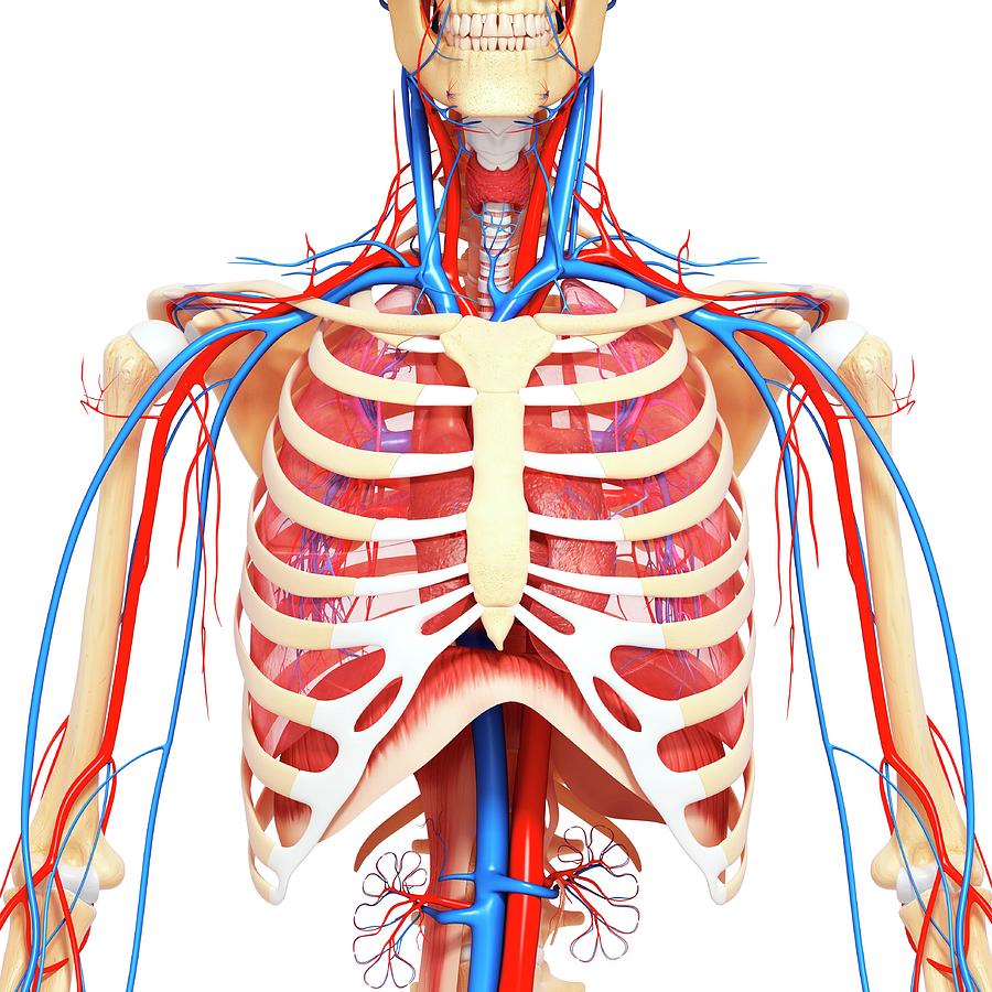
Medical illustration of circulatory system with heart and veins visible.
Chester chest with peripheral port access arm. These lungpatterns will discussed in more detail in an article. Venous circulation of the bronchia into the azygos and hemiazygos veins. Learn about each muscle, their locations & functional anatomy. Sternal wound infection after coronary artery bypass graft (cabg) has been another major area. In fact every radiologist and pulmonary physician should be an expert in chest film reading. Profile view of female chest area. Radiology basics of chest ct anatomy with annotated coronal images and scrollable axial images to help medical students and junior doctors learning anatomy. Learn about chest anatomy with free interactive flashcards. 1, inferior lobe of right lung. Diagrams of normal venous anatomy of the thorax. Chest, abdomen, pelvisprovides detailed views of anatomic structures in general anatomy and function chest wall. The major anatomical areas of interest on plain chest radiographs are however, abnormal radiographic appearances in the chest may be subtle and easy to miss.
These lungpatterns will discussed in more detail in an article anatomy of chest. Here's a useful infographic to help you learn about the abs.


0 Komentar